
Teleradiology
Sports-Related Injuries and the Importance of Radiology
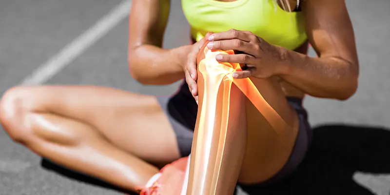
Almost one-third of all injuries faced by professional athletes are sports-related injuries. They also result in time lost to competition. Balancing the need to prevent injury from worsening or recurring and the desire to return the athlete to the competition remains the key objective of a sports medicine physician. Imaging provides a crucial link to confirm and assess the nature of the injury to guide the next steps in management and prognosis. Hence, to provide a proper diagnosis, we need accurate imaging of the body which signifies the importance of radiology in sports-related Injuries. When the grade or diagnosis of injury lacks clarity, the path to recovery remains uncertain. Ultrasonography and magnetic resonance imaging find wide application in sports injury and guide the need for interventional or surgical management.
Mechanism of Injury and Presentation
Indirect muscle injury or muscle strain is most commonly seen in elite athletes. During an eccentric contraction, there can be muscle disruption as the active contraction is added to passive stretching at the myotendinous junction (MTJ). Acute muscle injury results from sprinting and stretching and mainly affects the hamstring muscle complex. The recovery is faster for sprinting-related injuries even though is associated with a pronounced loss of function. Stretching injuries are associated with less loss of function and also slower recovery. A few of the factors are known to be associated with most of the injuries include, eccentric contraction, muscles with fast twitch type 2 fibers, sudden change in muscle function, muscles crossing multiple joints (biceps femoris, rectus femoris, gastrocnemius), failure to counteract forces from other muscles or ground reaction, and muscle Imbalance.
Indirect mechanisms have the propensity to acute avulsion injuries owing to extreme, unbalanced, and eccentric forces. Muscle strain can be categorised clinically into grade 1 with no appreciable tissue tearing, grade 2, tissue damage of MTJ with reduced strength and some residual function, and grade 3, a complete tear of MTJ unit with complete loss of function and occasionally a palpable gap. Direct muscle injury is usually caused by blunt trauma, mainly caused by collisions in soccer, football, and rugby. Massive blunt injury directed towards the bone can cause high-grade injury.
Muscle injuries are classified based as follows:
Grade I- Low disability, localised pain, small haemorrhage, swelling, and <10% ROM limitation
Grade II- Moderate disability, pain, and swelling, 5-50% loss of function, 10-25% ROM limitation
Grade III -Muscle rupture with severe disability and pain, loss of function of more than 50%, and severe ROM limitation of up to 25% Muscle contusion can clinically be categorised as mild when the range of a loss of motion is less than 1/3 rd with shorter recovery, moderate (ROM loss is less than 1/3 rd to 2/3 rd ) with moderate recovery times, and severe contusions (ROM loss greater than 2/3 rd ) with longer recovery times. Muscle strain is associated with more symptoms compared to that due to contusion.
Assessment of skeletal muscle injuries
Ultrasound (USG) advances enable visualisation of muscular architecture at the in-plane resolution, under 200 μm and with a section thickness of 0.5–1.0 mm and therefore better than MRI. USG being relatively inexpensive, and easier for patients, allow the serial evaluation to follow healing and can be performed in real time. The importance of radiology lies in its ability to diagnose medical conditions effectively through the use of advanced imaging technologies. USG has the additional advantage of showing muscle structure and surrounding anatomy that does not get obscured as in an MRI.
Clinical history can guide physical examination and subsequent USG. The use of a linear transducer can easily identify skeletal muscles. Multifrequency transducers allow visualisation of most muscle groups, whereas lower frequency linear probes may be needed in very muscular patients (gluteal and proximal thigh). Trapezoid fields of view (FOV) to 60cm with the help of composite image formation are enabled by modern software and transducers. Once optimal transducer setting is achieved, the symptomatic area can be scanned longitudinally and transversely. Initial assessment should be done at rest and then the surrounding tissues should be assessed with the help of active and or passive contraction to evaluate the consistency of abnormality (solid or cyst), alteration in muscle function, and any movement of disrupted fibers (grade or tears). Certain hernias may become apparent only on standing.
Normally the muscles are arranged in parallel hypoechoic bundles surrounded by echogenic fibrofatty septa. Adjacent fascia and nervous tissue are of higher echogenicity. Perimysium is relatively echogenic and can be viewed as parallel lines forming oblique angles with MTJ. The orientation of the perimysium is oblique in uni-bipennate muscles and parallel in fusiform muscles.
The transverse plane shows muscle fibers as hypoechoic with intervening septae seen as smaller linear areas and echogenic dots. Epimysium surrounding the entire muscle is echogenic due to its fibrous nature. The following grading is enabled by USG.
Grade 1 - Focal or diffuse ill-defined area of increased echogenicity within the muscle at the site of injury or no abnormality. There may be minimal focal fiber disruption occupying less than 5% of the cross-sectional area of the muscle, which is a well-defined focal hypoechoic or anechoic area within the muscle.
Grade 2 - Partial fiber disruption of less than 100% of the cross-sectional area of muscle affected. Discontinuity of echogenic perimysial stria around the MT. Intramuscular hematoma may be apparent within 24-48 hrs of injury as an ill-defined muscle laceration separated by hypoechoic fluid with marked increased reflectivity in the surrounding muscle. Post 48-72 hours, the well-defined hypoechoic fluid collection with echogenic margin gradually enlarges filling the hematoma in a centripetal manner.
Grade 3 - There may be a complete discontinuity or disruption of the MT with different degrees of retraction. There may be a palpable gap between the retracted ends of the muscles affected. The perifascial fluid which is usually hypoechoic may have increased echogenicity due to extravascular blood after 24 hours. However, the detection of perifascial fluid is not a specific feature to grade injury Pitfalls of USG evaluation of muscle injury Septae have linear configurations and are susceptible to anisotropy artifacts leading to decreased echogenicity and can be mistaken for an injury. Probe repositioning can help to rule out that the apparent absence of septae is due to injury or artifact. Prominent intramuscular vessels can mimic tears. This can be avoided by the use of Doppler and tracing the vessels through the septae to their neurovascular bundles and determining if the surrounding muscle structure is normal right up to the vessel boundary. Thickened and scarred septae can cause acoustic hypoechoic shadowing. Blood flow through muscles and connective tissues can increase during exercise by 20-fold, resulting in a 10-15% increase in muscle volume.
Routine MRI assessment of skeletal muscles
MRI is better suited when the injury is deep involving deep muscle compartments. Owing to its ability to visualise soft tissues with excellent contrast and provide high spatial resolution and multi-planar assessment MRI is the reference option when the morphology of muscles in athletes is needed to be evaluated. Therefore, is well suited to confirm and evaluate the extent and severity of muscle injuries. The growth of the healthcare sector has led to an increase in the number of medical imaging companies in India offering a wide range of imaging services. In order to ensure high-resolution images with thinner sections and smaller FOV, MR imaging is performed unilaterally by using a dedicated surface coil. When the bilateral injury is suspected, simultaneous acquisition of images of the contralateral lower limb by using higher FOV.
Based on the FOV desired, coil selection should be planned. Skin markers over the area of symptoms can help to correlate imaging with clinical features. Multiplanar (axial, coronal, and sagittal) acquisitions can accurately evaluate the morphology and extent of muscle injuries. Edematous changes around the MTJ and myofascial junctions can be detected by pulse sequences that include fat-suppressed fluid-sensitive techniques. Fluid-sensitive techniques include fat-suppressed spin-echo T2-weighted, proton density-weighted sequences, short-tau inversion recovery, or STIR technique. However, T1-weighted spin-echo sequences are less sensitive to muscle edema after acute injury. Most commonly the injuries occur around MTJ and edema and blood collection extend along the adjacent muscle, fibers, and fascicles. This may be detected on fluid-sensitive coronal or sagittal MR images as an ill-defined focal or diffuse high-signal intensity area along the MTJ is seen on images obtained with fluid-sensitive techniques with a classic feathery appearance.
In the year 2013, a consensus statement on new terminology and classification of muscle injuries in sports was published. Based on the cause of injuries the grading was defined.
Grade 1 - Fatigue induced disorders and delayed onset muscle soreness (DOMS)
Grade 2 - Spine related and muscle-related neuromuscular disorder
Grade 3 - Indirect injury mechanism that may include partial muscle tear
Grade 4 - Subtotal/complete discontinuity of muscle/tendon
Direct injuries or lacerations are not part of this consensus. DOMS is an overuse injury, where pain may develop hours or days after a specific activity. There may be decreased muscle function and strength. Pain peaks in 24 to 72 hours after physical activity and then decreases slowly. Clinically strain is characterised by the immediate onset of pain and hence can be easily differentiated. However, an MRI may be hard to distinguish. There is never a tear but may display diffuse edema affecting the muscle belly without the typical “feathery” pattern of strain and no perifascial fluid. There may not be any linear relationship between signal intensity on T2 weighted images and clinical symptoms. The symptoms may resolve within 10-12 days, whereas abnormal signal intensity on fluid-sensitive MRI may last up to 80 days.
Chronic exertional compartment syndrome (CECS) is characterised by chronic and recurrent pain induced by physical activity in young athletes owing to an abnormal increase of tissue pressures causing reduced perfusion and ischaemic pain. Intramuscular pressure before and immediately after exercise is recorded with a transducer to diagnose CECS. More than 20% change in T2 values in muscles before and after exercise is diagnostic of CECS.
Pathophysiology of muscle healing
The initial phase of healing is the destruction and inflammatory phase, followed by the repair phase around days 2 and 3 when both phagocytic necrosis and connective tissue scar accompanied by capillary ingrowth with the regeneration of skeletal muscles takes place. The third phase is remodeling which overlaps the repair phase.
Postinjury assessment
MRI shows fluid-like signal intensity at the site of injury. Scar tissue can be noted as early as 6 weeks after the initial injury and may appear as low signal intensity on T1 weighted and high signal intensity on fluid-sensitive MRI at early stages. Scar tissues display low signal intensity for all MRI pulse sequences. Residual scarring may be a common cause of misdiagnosis and also alter the mechanism of contraction during movement and increase the risk of re-injury. MRI changes may be seen even after the resolution of clinical symptoms.
USG of up to 50% of cases of grade 1 injuries appear with increased echogenicity. Therefore, healing appears as a reduction in the size or resolution of this increased echogenicity. Grade 2 injuries may appear to be hypoechoic owing to fluid adjacent to muscle fibrils pr epimysium.
Healing shows up as a substantial decrease in the quantity of fluid. Macroscopic muscle tears may show echogenicity of the margins of the tear with healing. USG may be less sensitive compared to MRI to evaluate residual morphological changes after muscle injury as MRI can offer higher soft tissue contrast and sensitivity to extracellular fluids. Dynamic assessment before and after muscle contraction is the major advantage associated with the use of USG. Follow-up may be relevant for grade 2 injuries with clinical symptoms even after proper management and rehabilitation and for reinjuries.
Complications may include hernia, myositis ossificans, compartment syndrome, and muscle atrophy, Elastosonography can allow real-time imaging of tissue elasticity. Following injury, there is increased elasticity owing to hematoma formation. Progressive resorption of hematoma and scar formation leads to loss of elasticity. An area with reduced elasticity extends beyond the US zone of the scar and has a worse prognosis.
Return to play and risk of reinjury
The choice for predicting the time to return in an elite-athlete setting to sports after acute muscle injury may be considered by some as MRI. However current literature may be limited to return to play after acute hamstring injuries. Multivariate statistical analysis confirmed that routine MRI cannot be recommended to provide an accurate return playtime. But plays a critical role in confirming the clinical diagnosis, and informing the athlete. There are only three studies published on MRI at a return to play following a hamstring injury that show persistently increased signal intensity on fluid-sensitive images at the return to sports clearance. Hence normalisation of signal intensity on fluid-sensitive images at a return to sport clearance may not be required for a successful return to play as functional recovery preceded structural recovery.
Reinjury risk remains high when there is a long absence from sports following the initial injury. Evaluation of MRI parameters in hamstring injury shows conflicting results on the predictive value of the risk of re-injury. However, confirmation of clinically suspected biceps femoris long head injury with MRI might be able to guide the clinician in predicting the risk of reinjury which is 16% compared to 2% for the medial injured group.
Conclusion
Loss of competition, long period of recovery, and risk of recurrent injury may be seen in both professional and amateur athletes owing to injury to the muscle. Optimal management can ensure adequate healing, low complications, and risk of recurrent injury. Ultrasonography is done by most medical imaging companies in India. USG being relatively cheap and fast allows for serial evaluation of the healing process and dynamic muscle assessment. It can guide the key elements to define recovery and rehabilitation.
More from AMI
Career in Radiology
29/08/2023
Empowering Radiologists-Teleradiology Redefines the Role of Imaging Specialist
09/10/2023
How to Choose a Prospective Teleradiology Service Provider
04/08/2023
Intra- Operative 3D Imaging With O- Arm Making Complex Spine Surgeries Safe and Accurate
30/11/-0001
Imaging Instrumentation Trends In Clinical Oncology
26/06/2023
Test
27/03/2024
Advances In Neuroradiology
06/01/2023
Emerging Techniques in Radiology By Dr. Namita
10/11/2022
Imaging In Pregnancy
18/01/2023
Behind The Scenes of Teleradiology: How Digital Imaging Is Changing Diagnostic Medicine
16/10/2023
Safe Radiology Practise for a safer world, safer health
30/11/2022
What is Diagnostic Radiology? Tests and Procedures
11/08/2023
Teleradiology's Contribution to Timely Emergency Diagnoses
04/10/2023
10 Strategies To Prevent Burnout In Radiology
09/01/2023
How to increase the efficiency of the Radiology Equipment
18/08/2023
Improvement of Patient Care Through Teleradiology
05/07/2023
Revolutionizing Indian Healthcare: Unlocking the Potential of Teleradiology in Remote Areas
27/09/2023

AMI Expertise - When You Need It, Where You Need It.
Partner With Us
